Speciality
Biometry
One of the most important tests performed prior to a cataract surgery. On which the surgery and its outcome is dependent. At AEC, we're result oriented. With advancement in technology, we upgrade ourselves. One of the upgradation at AEC is the state of the art IOL MASTER 700. Manufactured by renowned Zeiss, is now present at renowned , akshar eye clinic. This equipment not only measures eyes but also gives a better idea about visual outcome. It provides high definition pictures of inner structures of eyes which helps us to return your vision back in high definition. And that's not it, the equipment is even faster and accurate and more comfortable for our patients. At least 6 parts of eyes are viewed in less than two minutes.
Visuscout 100
Computers became laptops , smart phone became smart watches. With all gadgets turning compact, even eye care cameras have become compact. The latest addition at AEC is the visuscout 100 which is a hand held fundus(retina) camera which enables us to capture HD images of your retina hassle free. Not only normal images but also special red free images, which helps us for better diagnosis of retinal conditions.
C3r
Keratoconous is a progressive and painless corneal weakening which has direct impact on the vision. And makes day to day life difficult for an individual.
It is a condition which cannot be cured but the progression can be stopped by making the cornea stronger. In order to do that, C3r is performed, in which, the cornea is made stronger by riboflavin (vitamin B 2) under the presence of UV light. Its just like cementing of a weak wall. Once the cornea is made stronger and controlling or stopping the progression, the patient can use Gas permeable contact lenses and enjoy their vision again.
Contact Lenses
The most common replacement for glasses are contact lenses, some use it for cosmetic reasons while some use in way for therapeutic. Whatever your reason is, we can help you with your demand for contact lenses. Be it coloured lenses for the party or a gas permeable lens for keratoconus, the optometrists at AEC are skillful to consult, advise and fit soft and gas permeable contact lenses. Right from advising the best choice of lenses for your eyes(which is a tailor made advise) to train you to handle lenses is performed. An extensive work up is done so that your experience with the contact lenses stays the best.
Retina
Retina, the power switch of your eyes, helps you to see. Evaluation of the retina needs very accurate visualization of different layers of retina, which is not possible by naked eyes. So here we are, equipped with the Swiss knife tool in retinal diagnostic, the optos OCT-SLO. Helps us to visualise your retina in a way like no other. We can diagnose a retinal disease, have HD images, along with diagnosis we can also track the progression or regression of the conditions as well. It can measure quality and quantity of vision on the micro points on the retina and never misses. Not only retina, it helps to to take HD images of the cornea, angles of eyes. This equipment takes care of visualization of front and back of your eyes and helps us to treat you more 3D images are acquired . Doesn't only diagnoses a condition but also helps to quantify it. Efficiently and accurately .
IOL
India is getting digital and modern day by day and so are our senior citizens. Every person has a wish, there should be no changes in day to day life after a cataract surgery, example:- using a laptop or a cell phone. The most frequently asked question by to-be cataract operated patients is "will I need glasses to use my cell phone or read my book after surgery?" Well we have an answer which you wish to hear "No need of glasses anymore!" Everyone has their own and different visual demands. We implant different IOLs based on your visual demands. So that your life would be uninterrupted but also your visual performance would increase. If you're a long term reader but don't want to wear glasses after surgery, here we are with the option of a multifocal IOL. It not only takes care or your far distance tasks but also near tasks, without glasses! If you're in a office set up where you have to use your laptop along with paper work, you can opt for the most modern IOL, that's a trifocal IOL. It takes care of far distance, near as well as intermediate distance, I.e the distance at which a laptop or computer is placed. On implantation of this kind of IOL, we take you back to your 20s when you never needed glasses for these kind of tasks. A matter of fact is these kind of implantation needs expertise, Dr nitin malkan, being the first surgeon to implant such an iol in Maharashtra holds a great experience in implanting such modern IOLs. If your retirement plan is to play a sport like golf or cricket and have High astigmatism, nothing to worry about, you wont be needing glasses anymore to play your sports. A toric IOL is the best option for you, correcting your astigmatism it provides the best quality and quantity of vision enabling you to be a champion of your desired sport. An office goer who doesnt want to wear glasses, but has astigmatism, well, a multifocal toric IOL is the answer. Which will correct far distance, near as well as astigmatism!
HFA 3
Visual fields, one of the most important tests performed to track the progression of glaucoma and also other neurological defects. Requires accuracy and speed. Based on it, AEC is happy to announce the addition of the latest version of Zeiss Humphrey field analyzer, which has one of the kind LIQUID LENS . Not just that, the equipment ensures speed, accuracy and efficacy like never before. And its the most advanced equipment to measure the visual fields till date. Helps us to serve you better .
Low Vision
What is low vision?
Low vision is a subspecialty within the professions of optometry and ophthalmology dealing with individuals who have reduced vision even when using the best possible spectacle or contact lens correction available. It can be a result of either congenital disease or acquired factors
Is low vision just part of getting older?
No. Some normal changes in our eyes and vision occur as we get older. However, these changes usually don't lead to low vision.
Most people develop low vision because of eye diseases and health conditions .While vision that's lost usually cannot be restored, many people can make the most of the vision they have
Your eye care professional can tell the difference between normal changes in the aging eye and those caused by eye diseases.
How do i know when to get eye exam?
Regular dilated eye exams should be part of your routine health care. However, if you believe your vision has recently changed, you should see your eye care professional as soon as possible.
Defination of low vision
Low vision is a significant reduction of visual function that cannot be fully corrected by ordinary glasses, contact lenses, medical treatment and/or surgery. Low vision affects people of all ages...in the home, on the job, and at school.
Low vision is a term that denotes a level of vision that is 20/70 or worse and cannot be fully corrected with conventional glasses. Low vision is not the same as blindness. Unlike a person who is blind, a person with low vision has some useful sight. However, low vision usually interferes with the performance of daily activities
Classifying low vision
Anyone with reduced vision not corrected by spectacles or contact lenses can be considered to be visually impaired. The World Health Organization uses the following classifications of visual impairment. When the vision in the better eye with best possible glasses correction is:
- 20/30 to 20/60 : is considered mild vision loss, or near-normal vision
- 20/70 to 20/160 : is considered moderate visual impairment, or moderate low vision
- 20/200 to 20/400 : is considered severe visual impairment, or severe low vision
- 20/500 to 20/1,000 : is considered profound visual impairment, or profound low vision
- less than 20/1,000 : is considered near-total visual impairment, or near total blindness
- No Light Perception : is considered total visual impairment, or total blindness
How do i know if i have low vision?
There are many signs that can signal vision loss. For example, even with your regular glasses, do you have difficulty-?
- Recognizing faces of friends and relatives?
- Doing things that require you to see well up close, like reading, cooking, sewing, or fixing things around the house?
- Picking out and matching the color of your clothes?
- Doing things at work or home because lights seem dimmer than they used to?
- Reading Street and bus signs or the names of stores?
Vision changes like these could be early warning signs of eye disease. Usually, the earlier your problem is diagnosed, the better the chance of successful treatment and keeping your remaining vision.
Magnitude of visual impairment
Globally, in 2002 more than 161 million people were visually impaired, of whom 124 million people had low vision and 37 million were blind. However, refractive error as a cause of visual impairment was not included, which implies that the actual global magnitude of visual impairment is greater
Symptomps of low vision
- Difficulty recognizing objects at a distance (street signs or bus signs)
- Difficulty differentiating colors (particularly in the green-blue-violet range)
- Difficulty seeing well up close (reading or cooking)
The symptoms described above may not necessarily mean that you have low vision. However, if you experience one or more of these symptoms, contact your eye doctor for a complete exam. Your eye doctor can tell the difference between normal changes which are common with age and changes caused by eye disease Top
Causes of low vision
Pathologies which may cause vision acuity loss
- macular degeneration (the most common cause of low vision; involves damage to a person's central vision making it difficult to read, drive, or perform other daily activities that require fine, central vision)
- Cataracts
- Glaucoma
- Uveitis
- Corneal opacity
- Trachoma
- Diabetic retinopathy
- Myopia magna
- Stargardt's disease
- Albinism
- Retinitis pigmentosa
- Amblyopia
- Aging (Aging is a risk factor for low vision; however, persons of any age may be affected.)
- Congenital defects (present at birth)
- Injury
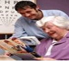
Low Vision Examination / Treatment
Vision and Visual Rehabilitative Services Clinic embraces a multi-disciplinary approach to the treatment of low vision. Ophthalmologists, optometrists and occupational therapists make up the team of health care professionals who will work with you starting with your vision examination, and continuing to work with you to identify treatment options, which include:
- Optical devices
- Techniques that will help you utilize your remaining vision.
- Environmental modifications to maximize your remaining vision.
- Adaptive non-optical devices,
- Occupational therapy programs may last as long as several months or be as brief as one session.
What Are Low Vision Devicies/AIDS
Because low vision cannot be improved by more traditional methods (i.e., the use of eyeglasses, contact lenses, etc.) persons with low vision often rely on the use of a number of different instruments, called low vision devices, and tailored equipment for improved vision. Low vision devices, categorized as either optical or non-optical, help to improve visual ability for millions of people everyday.
TopWhat Are Optical Low Visual AIDS
Simply stated, optical low vision devices involve the use of one of many types of lenses to improve vision. For example: Low visual aids can be categorized-
- Glasses- Eyeglasses are not considered a low vision device unless they have a high reading "add" or contrast enhancement tint, but they deserve special mention here since glasses ensure that the person's eyes see the clearest image possible and can easily focus at close distances
- Contrast enhancement aids- it includes different types of filters, dark felt tip ball pens etc
- Near visual aids- it includes dome magnifier, hand held magnifiers, illuminated hand held magnifiers, prismo half eye glasses, half eye glasses with high adds, pocket magnifiers, stand magnifier, etc
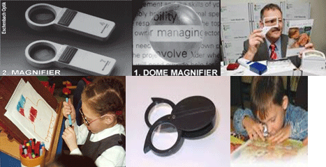
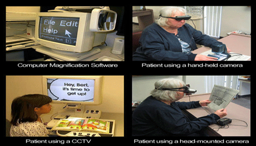
- Computers and software - Desktop and laptop computers have advanced the ability. Magnification can be changed in common software such as Microsoft Outlook, Excel, and Word. Specialized software is also available
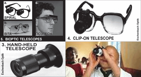
- Distance visual aids- it includes different types of hand held telescopes, clip-on telescopes, bioptic telescopes
- Electronic aids- it includes closed circuit television , electronic stand magnifiers, magnicam
Effectively of optical devices-
Adaptation process to visual aids In the patient's first visit, the most adequate options for their particular case are studied, taking into consideration their psychological, cultural, social and work factors, and the degree of improvement experienced with the selected aids, advising the patient on which aids will yield a better quality of life.
What are non optical low visual AIDS
Non-optical low vision devices help bring images closer to the eyes. This may include the use of any, or all, of the following:
- larger print items (i.e., magazines, newspapers, books, calendars, address books, cookbooks, dictionaries, games, playing cards, sheet music, street signs, etc.)
- larger, illuminated watches and clocks
- writing guides
- Typoscopes
- Notex
- signature guide
- Cheque guide
- Instruments that provide voice instruction (i.e., computers)
- Instruments that provide voice information (i.e., clocks, timers, calculators, scales, key chains, etc.)
Other Aids
For the totally blind, there are books in Braille, audio-books, machines and computer programs which transform text files into sound. Low vision people can, of course, make use of these tools as well. Computers are, precisely, fundamental tools of integration for the visually impaired person. They allow, using standard or specific programs, screen magnification and conversion of text into sound or touch (Braille line), and are useful for all levels of visual handicap
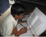
Low vision Therapy/ Training
Low vision therapy is any individual tailored tuition in the use of vision or low visual aids
What is Low Vision Therapy
Top- It teaches people how to use low visual devices
- Training in the adaptation and use of the environment
- Transfers skills from hospital to home
- Teaches about 3b,s, how to make things big, bold and bright
Conclusion
An ever-increasing number of people are at risk of visual impairment as populations grow and demographic shifts move towards the predominance of older age groups. Potentially blinding eye conditions such as age-related macular degeneration (AMD), diabetic retinopathy and glaucoma are increasing as the number of people affected grows. These are non-communicable chronic eye diseases to which the principles of long-term care including issues of cost of treatment and compliance (adherence) apply. We believe that low vision, as a complementary technique to ophthalmology, has a great future, due to the progress of science, the increase of life expectancy, and the increasing need people have to access information.
Where To Get Low Vision Work Up Or Training
At AKSHAR EYE CLINIC we have special trained optometrist for vision therapy and low vision assessment with special equipped and designed low vision devices with all favorable enviourment for low vision work up.
We examine the low vision patient, train them for how to use devices, rehabilitate, and dispense the devices as per the need of patient.
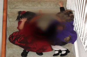
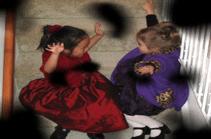
WHAT PEOPLE WITH LOW VISION SEE
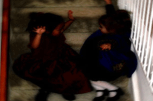
- Normal vision - This picture shows two children playing on a staircase

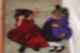
- Central field loss - A hazy or dark hole appears in the center of objects. Causes include macular degeneration and optic atrophy
- Multiple field loss- Scattered dark patches or holes appear around objects. Causes include diabetic retinopathy, glaucoma, retinal detachment and trauma.
- Tunnel vision - Loss of peripheral vision causes a restricted field of vision. Objects in the center remain visible. Causes include glaucoma, retinitis pigmentosa and stroke

- Contrast loss and glare problem- Objects blend in with the background; lights are distracting or uncomfortable. Causes include cataracts, glaucoma, corneal disease and albinism.
- Some additional symptoms may include- Blurred vision .Objects appear out of focus. Causes include macular degeneration, diabetic retinopathy, cataracts or corneal disease.
- Distortion- Objects appear crooked, wavy or doubled. Causes include macular degeneration, diabetic retinopathy and retinal detachment.

Top

Cataract
A cataract is a clouding that develops in the crystalline lens of the eye or in its envelope, varying in degree from slight to complete opacity and obstructing the passage of light.Although cataracts have no scientifically proven prevention, it is sometimes said that wearing ultraviolet-protecting sunglasses may slow the development of cataracts.
It is removed the most advanced micro co axial phacoemulsification with OZIL Infinity machine and using a top in the line Lumira Zeiss microscope.
Glaucoma
Glaucoma refers to a group of diseases that affect the optic nerve. It also involves a loss of retinal ganglion cells in a characteristic pattern. It is a type of optic neuropathy.Glaucoma can be divided roughly into two main categories, "open angle" and "closed angle" glaucoma. Closed angle glaucoma can appear suddenly and is often painful; visual loss can progress quickly but the discomfort often leads patients to seek medical attention before permanent damage occurs. Open angle, chronic glaucoma tends to progress more slowly and the patient may not notice that they have lost vision until the disease has progressed significantly.
Our centre is more then fully equipped to detect earliest damage due to Glaucoma with the help of SWAP(short wave automated perimetry), GPA(glaucoma progression analysis) software and RNFL(retinal nerve fibre layer) analysis by spectral domain OTI OCT machine. Each and every patient is routinely screened to rule out glaucoma.
Cornea
It is the transparent front part of the eye that covers the iris, pupil, and anterior chamber. Together with the lens, the cornea refracts light, accounting for approximately two-thirds of the eye's total optical power.Various refractive eye surgery techniques change the shape of the cornea in order to reduce the need for corrective lenses or otherwise improve the refractive state of the eye. In many of the techniques used today, reshaping of the cornea is performed by photoablation using the excimer laser.
Our clinic is equipped with state of the art AXIS 9000 Zeiss corneal topography machine to detect various corneal disorders and Tomy corneal Pachymeter for measuring corneal thickness.Our clinic is equipped with state of the art AXIS 9000 Zeiss corneal topography machine to detect various corneal disorders and Tomy corneal Pachymeter for measuring corneal thickness.
Oculoplasty
Oculoplastic Surgery, also known as Ophthalmic Plastic and Reconstructive, Oculofacial or Eye Plastic Surgery, is a surgical subspecialty of Ophthalmology that deals with the medical and surgical management of deformities and abnormalities of the eyelids, lacrimal (tear) system, orbit and the adjacent face (cosmetic lid and brow surgeries).Occuloplasty surgical division treats drooping of the eyelids, all types of eyelid abnormalities and Botox treatment for various skin conditions around the eye.
TopPediatric Ophthalmology
Pediatric ophthalmologists focus on the development of the visual system and the various diseases that disrupt visual development in children. Pediatric ophthalmologists also have expertise in managing the various ocular diseases that affect children. Pediatric ophthalmologists are qualified to perform complex eye surgery as well as to manage children's eye problems using glasses and medications.The department runs regular amblyopia clinic and squint clinic. The department runs regular amblyopia clinic and squint clinic.Diabetic Retinopathy
Diabetic retinopathy is retinopathy (damage to the retina) caused by complications of diabetes mellitus, which can eventually lead to blindness. It is an ocular manifestation of systemic disease which affects up to 80% of all patients who have had diabetes for 10 years or more. Despite these intimidating statistics, research indicates that at least 90% of these new cases could be reduced if there was proper and vigilant treatment and monitoring of the eyesOur clinic organises Diabetes awareness programmes for the patients with the help of Retinal surgeon, Pathologist and Diabetologist. The clinic is equipped with angiography, laser treatment and a full fledged pathology laboratory for diabetic patients. The clinic is regularly attended by a diabetologist.
Retina
The vertebrate retina is a light sensitive tissue lining the inner surface of the eye. The optics of the eye create an image of the visual world on the retina, which serves much the same function as the film in a camera. Light striking the retina initiates a cascade of chemical and electrical events that ultimately trigger nerve impulses.A number of different instruments are available for the diagnosis of diseases and disorders affecting the retina. An ophthalmoscope is used to examine the retina. Recently, adaptive optics has been used to image individual rods and cones in the living human retina and a company based in Scotland have engineered technology that allows physicians to observe the complete retina without any discomfort to patients
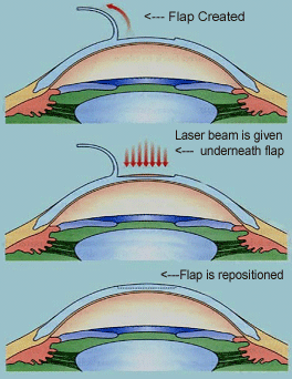
LASIK
LASIK (laser assisted in situ keratomileusis)There are different ways to correct refractive errors like glasses, contact lens and refractive surgery
Lasik is a type of refractive surgery to correct the refractive error
Technology
Tecnonology has emerged like anything, Lasik is completely different procedure than early days refractive surgery like RK (radial Keratotomy),or PRK( photorefractive keratectomy) as it provides immediate improvements in vision and involves much less pain and discomfort
TopRange (Scopes)
1 to 20 diopters of myopia (depending on corneal thickness)
1 to 8 diopters of hypermetropia
Up to 8 diopters of astigmatism
Pre - Operative Evaluation
- Complete eye examination by ophthalmologist
- Age -18 years and above
- Refractive error or refraction should be stable
- Central Corneal thickness should be measured to decide whether is it enough for corneal ablation using Pachymeter
- Corneal mapping using corneal topographer to understand anatomy of each area on cornea
- Contact lens wearer are instructed to stop wearing 7days prior to Lasik in case of soft contact lens and 6 weeks prior for hard or semisoft lenses
- An anti-biotic eye drop is prescribed 2 days prior to the surgery to avoid the risk of infections
Procedure
Generally a mild sedative and anesthetic eye drops are instilled before the procedure LASIK is performed in three steps- The first step is to create the flap of corneal tissue
- Second step is remodeling the cornea under the flap with laser
- Finally the flap is repositioned
Post Operative Care
- Generally 2 days rest is advised and proper instillations of drops as prescribed
- Antibiotic drops are prescribed fro few days
- Anti inflammatory drops are prescribed post operatively for few days
- Lubricating drops are advised
- Darkened pair of shields are prescribed to reduce glare and rubbing of eyes for few days
- Gymnastic exercises is to be avoided for atleast 1 month
- Swimming is to be avoided for atleast 15 days
Post –Operative Complications
3-6 % patients suffer from some complications within the span of 6 months
Immediate complications which are resolved are more common- Sub-conjunctival hemorrhage
- Dry eye
- Over and under correction
- DLK (diffuse lamellar keratitis - Sands of Sahara syndrome)
- Visual disturbances (fluctuation in vision, startbursts, halos, glare, photophobia (light sensitivity), double vision, ghost images
- Flap striae
- Epithelial ingrowth under the flap
- Displaced flap
- Corneal infections
- Corneal ectasia
- Macular hole
Also Visit


News & Events
Akshar eye clinic proudly acounces the addition of new machine that is Zeiss FDT( Frequency doubling technolgy )Perimetry. The instrument is designed for fast and effective detection of visual loss. Which one of the few ophthalmologist have.

Seasonal Disease
Allergic conjunctivitis or spring catarrh is a very common seasonal disorder in children between 5 - 15 years of age. The disorder can only be suppressed. Cure cannot be offered.
Download Brochure