

About Eye-Anatomy Of The Eye
Human Eye Anatomy
THE HUMAN EYE
The eye allows the perception and interpretation of the shapes, colors, and dimension of objects in the world by processing the light they reflect. The eye can see in bright or dim light but not when light is absent.
PROCESS OF VISION
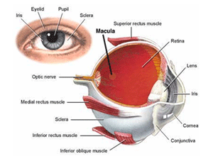
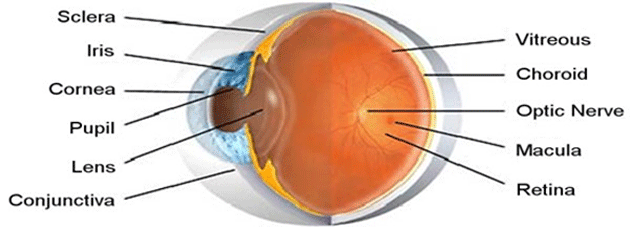
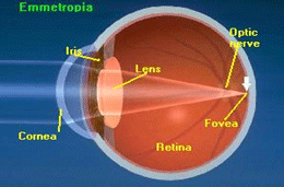
Light waves from an object ( such as a tree) enter the eye first through the cornea, which is the clear dome in front of the eye, then progresses through the pupil, the circular opening in the center of the colored iris, then passes through the lens, located immediately behind the iris and the pupil. The light continues through the vitreous humor, the clear gel that makes up about 80% of the eyes volume, and then, ideally back to a clear focus on the retina behind the vitreous. The small central area of the retina is Macula, which provides the best vision of any location in the retina. Initially, the light waves are bent or converged first by the cornea, and then further by the crystalline lens. At that point, the image becomes reversed (turned backwards) and inverted (turned upside-down).
EYE STRUCTURES
The most important components of the human eye are the Cornea, Conjunctiva, Iris, Crystalline lens, Vitreous Humor, Retina, Macula, Optic, Nerve, and Extra Ocular Muscles.
ORBIT
The eyeball is set in a protective cone-shaped cavity in the skull called the orbit or socket.
SCLERA
Along its circumference, the cornea is continuous with the sclera: the white, opaque portion of the eye. The sclera makes up the five- sixth of the eyes outer layer. It provides protection and serves as an attachment for the Extra Ocular Muscles which moves the eye.
Disorders of Sclera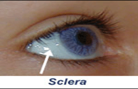
- Scleritis
- Episcleritis
CONJUNCTIVA
The conjunctiva is mucous membrane that lines the inner surfaces of the eyelids and folds back to cover the front surface of the eyeball, except for the central clear portion of the outer eye (the cornea).
Disorders of conjunctiva
- Conjunctivitis
- Conjunctival cyst
- Pterygium
- Pinguincula
TEAR GLANDS
Coating the outer surface of the cornea is a pre-corneal tear film. People normally blink the eyelids of their eyes about every six seconds to replenish the tear film.
Tears have four main functions on the eye:
- Wetting the eye.
- Creating a smooth optical surface.
- Acting as the main supplier of oxygen and other nutrients to the cornea.
- Containing an enzyme to destroy bacteria.
CORNEA
One-sixth of the outer layer of the eye bulges forward as the cornea, the transparent dome which serves as the outer window of the eye. The cornea is the primary (most powerful) structure focusing light entering the eye (along with the secondary focusing structure, the crystalline lens).The net refractive power of the cornea is about +43.00 D which is 3\4th of the total power of the eye (60 Diopter). It is composed, for the most part, of connective tissue with a thin layer of epithelium on the surface.
Disorders of Cornea
- Keratitis
- Corneal Ulcers
- Keratoconus
- Corneal Opacities
IRIS
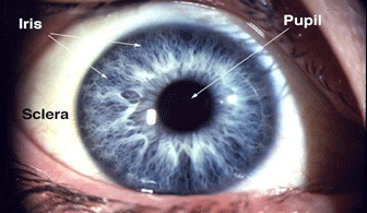 The iris is visible through the clear cornea as the colored disc inside the eye. It lies between the cornea and the crystalline lens. This tissue gives color to the eye.
PUPIL
The iris acts like the shutter of a camera. The pupil is a circular hole in the middle of the iris that regulates the amount of light passing through the retina. The pupil constricts and dilates to regulate light
LENS
The lens is a, transparent, Avascular, biconvex crystalline structure located in posterior chamber behind pupil and held in position by zonules, a membrane-like structure that is quite elastic. The refractive power of the lens is about 20 diopters.
Disorders of Lens:
- Cataract
- Subluxation of lens
RETINA:
It is the innermost sensory layer of an eye. Entire retina is 72% of a sphere about 22 mm in diameter. Retina contains nerve fibre cells.
Disorders of Retina:-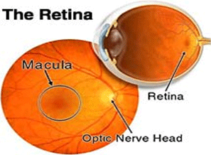
- ARMD
- Retinitis Pigmentosa
- Cystoid Macular Edema
- Central Serous Retinopathy
- CRAO
- CRVO
- BRVO
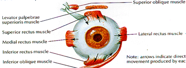
EXTRA OCULAR MUSCLES
The orbit is surrounded by layers of soft, fatty tissue which protect the eye and enable it to turn easily. Three pairs of extra ocular muscles regulate the motion of each eye.
Also Visit


News & Events
Akshar eye clinic proudly acounces the addition of new machine that is Zeiss FDT( Frequency doubling technolgy )Perimetry. The instrument is designed for fast and effective detection of visual loss. Which one of the few ophthalmologist have.

Seasonal Disease
Allergic conjunctivitis or spring catarrh is a very common seasonal disorder in children between 5 - 15 years of age. The disorder can only be suppressed. Cure cannot be offered.