About Eye - Keratoconus
KERATOCONUS
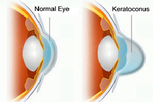
Keratoconus (kerato –horn, cornea & konos-cone) is a degenerative disorder of the eye in which structural changes within the cornea cause it to thin and change to a more conical shape than its normal gradual curve . As keratoconus progresses, the cornea bulges and thins, becoming irregular. The progression of KC is unpredictable. It is generally slow and can stop at any stage from mild to severe.

Etiology (Causes) of Keratoconus
The cause is unknown but the tendency to develop keratoconus is probably present from birth.Keratoconus is thought to involve a defect in collagen, the tissue that makes up most of the cornea .There is also systemic& an ocular associations.
Systemic disorder include Down,Turner,Ehlers-Danols & Marfan syndromes, atopy, osteogenesis imperfecta, mitral valve prolapsed & mental retardation.
Ocular associations include vernal keratoconjunctivitis, blue sclera, aniridia, ectopia lentis, leber congenital amaurosis, retinitis pigmentosa & persistent eye rubbing.
What are the symptoms of Keratoconus?
- The typical patient with undiagnosed keratoconus complains of deteriorating vision, usually in one eye first, both at distance and near.
- Near visual acuity may improve if the patient squints or holds printed material closer.
- Keratoconus patients often report multiple images or ghosting of images and often relate a history of frequent refractive correction changes without much improvement in visual acuity.
- Patients may also report irritating symptoms such as intolerance to glare, photophobia and a recurrent foreign body sensation.
- Even with appropriate contact lens correction, keratoconus patients often report fluctuating vision throughout the day and from day to day, although the measurements of visual acuity in keratoconus patients are highly repeatable.
Exams and Tests
- Keratoconus is frequently discovered during adolescence. It can usually be diagnosed with slit-lamp examination of the cornea. Early cases may require a test called corneal topography, which creates a map of the curvature of the cornea.
- When keratoconus is advanced, the cornea may be thinner in areas. This can be measured with a painless test called pachymetry

Treatment
- Spectacles in early cases to correct regular & mild irregular astigmatism.
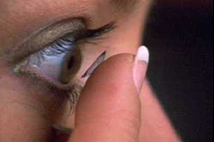
- Contact lenses: In early stages of keratoconus, soft contact lenses can suffice to correct for the mild astigmatism. As the condition progresses, these may no longer provide the patient with a satisfactory degree of visual acuity, and most clinical practitioners will move to managing the condition with rigid contact lenses, known as rigid gas-permeable, or RGPs. RGP lenses provide a good level of visual correction, but do not arrest progression of the condition.
- Some patients also find good vision correction and comfort with a "piggyback" lens combination, in which gas-permeable rigid lenses are worn over soft lenses, both providing a degree of vision correction One form of piggyback lens makes use of a soft lens with a countersunk central area to accept the rigid lens.
- Scleral lenses are sometimes prescribed for cases of advanced or very irregular keratoconus; these lenses cover a greater proportion of the surface of the eye and hence can offer improved stability. The larger size of the lenses may make them unappealing or uncomfortable to some; however, their easier handling can find favour with patients with reduced dexterity, such as the elderly
- Surgical options: Corneal collagen cross linking with riboflavin (C3R) Corneal cross linking is the newest development in the treatment of keratoconus and as such it holds a lot of promise. Quite simply, C3R involves treating the cornea with riboflavin and then activating its collagen cross linking properties with ultraviolet light. C3R improves the strength of the cornea by increasing the linking between collagen fibers. The result of this increased cross linking is that it increases the strength of the cornea and often causes some corneal flatten.A one-time application of riboflavin solution is administered to the eye and is activated by illumination with UV-A light for approximately 30 minutes. The riboflavin causes new bonds to form across adjacent collagen strands in the stromal layer of the cornea, which recovers and preserves some of the cornea's mechanical strength. The corneal epithelial layer is generally removed in order to increase penetration of the riboflavin into the stroma.
- Corneal ring segment inserts: A recent surgical alternative to corneal transplant is the insertion of intrastromal corneal ring segments. A small incision is made in the periphery of the cornea and two thin arcs of polymethyl metha acrylate are slid between the layers of the stroma on either side of the pupil before the incision is closed. The segments push out against the curvature of the cornea, flattening the peak of the cone and returning it to a more natural shape. The procedure, carried out on an outpatient basis under local anaesthesia, offers the benefit of being reversible and even potentially exchangeable as it involves no removal of eye tissue.
- keratoplasty
KERATOCONOUS MYTHS & FACTS
What is Keratoconous?
It is a unilateral/bilateral corneal condition where corneal shape is altered. In simple terms cornea (the black part of the eye) becomes conical. This results in frequent charge of glasses with high cylinder power. Despite wearing glasses patients may not have good visual acuity in this condition.
How does one get kerotoconous?
Kerotoconus etiology is still unknown. A number of factors have been postulated to be associated with kerotoconus. Genetic cause is a major one. Frequent eye-rubbing has also been postulated as an important cause of kerotoconus-this condition is also associated with a number of ocular and systemic conditions such as Retinitis Pigmentosa, allergic eye disease, Down's syndrome etc.
How does one get kerotoconous?
Most often kerotoconous is diagnosed in healthy young patients who go for refraction for change of glasses or incidentally noted when patients go for Lasik evaluation. Early symptoms are intolerance to glasses & frequent change of numbers. Poor vision with glasses. Early signs are a scessor reflex on retinoscopy. Advanced cases may be picked up by the classical protusion of cornea early cases are picked up using corneal topography test.
How does one get kerotoconous?
Keratoconous is a progressive condition: - the treatment options vary with the grade of the disease for early cases glasses may improve visual acuity. For moderate advanced cases contact lenses (RGP/ Rose K/Scleral lenses are good potion). For very advanced cases corneal transplantation may be required it is important to follow up every 3-6 months to rule out progressive keratoconous affects however it does not lead to blindness with proper optical correction visual acuity can be restored.
How does one get kerotoconous?
Yes with advent of technology like corneal collagen cross linking the progression of keratoconus can be halted. It also leads to a little improvement in the visual function
Can I Undergo Lasik
Lasik is usually contraindicated in kertoconous however with recent advance for low moderate kerotoconous we can how perform PRK with collagen crosslinking, Intact with corsslinking on even phakic IOL with collagen crosslinking make people free of glasses.
For Further quires please feel free to contact us at : akshareyeclinic@gmail.com
Phone No : 28080469/28080406
News & Events
Akshar eye clinic proudly acounces the addition of new machine that is Zeiss FDT( Frequency doubling technolgy )Perimetry. The instrument is designed for fast and effective detection of visual loss. Which one of the few ophthalmologist have.
Seasonal Disease
Allergic conjunctivitis or spring catarrh is a very common seasonal disorder in children between 5 - 15 years of age. The disorder can only be suppressed. Cure cannot be offered.Download Brochure


